Radiology

0 readers
1 users here now

A community for all things related to medical imaging!

Rules:

  1. Please be courteous and professional.
  2. No requests for medical advice or second opinions. Please see your actual physician/provider for that!
  3. No advertising.
  4. Imaging/case submissions must have no patient identifiers.
  5. Imaging/case submissions should have a known finding or point of interest.

founded 1 year ago
MODERATORS
1


54 year old female with a history of minor injury to the finger 6 months before these studies were obtained, subsequently developed an infection requiring debridement. The wound was then stable until 2 months before, when a large, fungating, hemorrhagic mass grew.

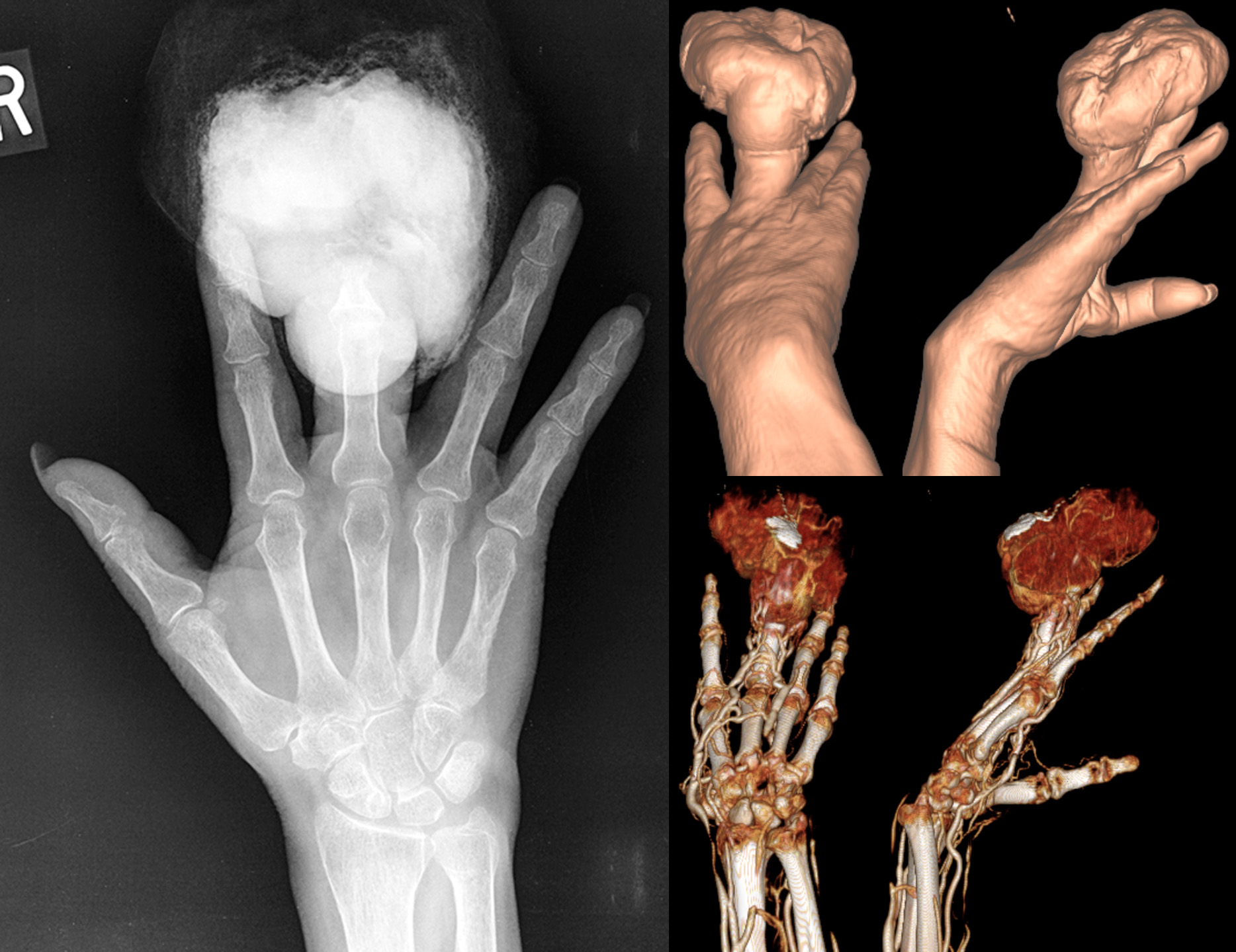
X-ray shows large radiopaque mass eroding the distal finger (distal phalanx and distal portion of the middle phalanx).

CT 3D surface reconstructions show the morphology of the mass.

CTA 3D reconstruction shows the mass is very hypervascular.

Lymph node scintigraphy was performed showing a sentinel node at the axilla (not shown). The patient underwent amputation of the finger with axillary sentinel node biopsy, which was positive for metastatic melanoma. The patient was then lost to follow-up.

2


38 year old guy fired his 45 mm into his knee while cleaning the gun. No exit wound.

There is a comminuted fracture of the distal femur with multiple bullet fragments, including the largest at the intercondylar notch. There is gas/air in the knee joint, forming air-fluid levels on lateral view. There are additional foci of gas in the surrounding soft tissues.

Patient underwent arthrotomy for foreign body removal and fixation of the supracondylar/intercondylar femoral fracture.

3


41 year old male with history of HIV-AIDS, meth-abuse, presents with progressive headache and confusion over days.

CT shows a large mass destroying the left frontoparietal calvarium and causing significant mass effect on the underlying brain.

MR shows a heterogeneous enhancing mass with hemorrhage (SWI - dark areas), hypercellularity (DWI - mildly bright areas), and hyperperfusion (ASL - bright areas), features suspicious for high-grade tumor.

4


This is my neck. I was born with this and it was discovered when I was 41. There are fused vertebrae from C4-C7, malformed discs, deformed vertebrae, a bulging disc touching my spinal cord and the dermoid tumour on my spinal cord.

5


62 year old female with a history of tobacco, opioid, and meth abuse as well as heart attack requiring coronary bypass and stents. She presents with bilateral lower extremity claudication (painful fatigue with physical activity) starting from her glutes.

CT angiogram shows occlusion of the lower aorta (red markings) as well as both common iliac arteries.

Vascular ultrasound shows very poor arterial waveforms throughout the lower extremities, essentially flat/absent below the knee.

She underwent a transabdominal aortobifemoral bypass to treat this.

Radiopedia article on Leriche syndrome.

Meth is bad.

6


Very unfortunate case of a young patient, who was crossing the street on a bike when he was struck by an SUV. The patient had brief loss of consciousness with decorticate posturing. Following that, he had reported no sensation and total weakness from the waist down, with physical exam showing total sensory and motor loss from the nipple level (T4) and below.

CT shows no fractures but some abnormal amount of gas/air in the back of the spinal canal at C7-T1.

MR shows complete transection of the spinal cord with 0.5 cm gap at T3 - goes well with the physical exam finding. There is a dorsal epidural hematoma. There was also disruption of the anterior longitudinal ligament, posterior longitudinal ligament, and ligamentum flavum at T3 (not shown).

What probably happened was that instead of a fracture, the traumatic force went through the ligaments maintaining spinal alignment, causing the spine at the T3 level to dislocate and shear, cutting through the spinal cord. By the time imaging was done, the bones relocated to their normal position, but the damage had been done.

7


Female in her 20s with (all right-sided) proptosis, intermittent vision loss, facial pain, and mid to lower face numbness.

Coronal and axial CT images show a homogenous bony expansile process that results in narrowing of the soft tissue compartments of the face. As a result, the right eye is pushed forward (proptosis; as seen on the MR image), and many of the right-sided (as well as some left-sided) skull base foramina that carry nerve bundles and blood vessels are severely narrowed.

8


Elderly female with a history of hypertension, stroke, and, 1 month before, heart attack with cardiac arrest. The patient presented to the emergency department with 1 day of nausea, vomiting, cold sweats, and malaise. Troponins 1.54. ECG with inferior Q waves but no ST elevation/depression or T-wave inversion.

Chest radiograph shows an enlarged cardiac silhouette, for which a differential of cardiomegaly or pericardial effusion was suggested, as well as increased pulmonary vascular markings compatible with pulmonary congestion. Compared chest radiograph findings to the images from the methamphetamine-induced cardiomyopathy case.

CT angiography shows both cardiomegaly as well as an intermediate-density pericardial effusion (magenta arrows) concerning for hemopericardium. Most concerningly (holy-shit concerningly), there is a left ventricular aneurysm with rupture and blush of contrast (red arrow) into the hemopericardium, compatible with ongoing bleeding.

It is not surprising that a rupture of the heart carries high mortality risk. The patient passed away from PEA cardiac arrest shortly after admission, likely from cardiac tamponade by the enlarging hemopericardium.

9


60 year old male with a history of homelessness, latent tuberculosis, and methamphetamine abuse. The patient was admitted to the hospital for shortness of breath and leg edema. A urine drug test on admission was positive for methamphetamine.

Chest radiograph (right) shows an enlarged cardiac silhouette, for which a differential of cardiomegaly or pericardial effusion was provided. Subsequent echocardiogram confirmed this was cardiomegaly with a low ejection fraction. The pulmonary vascular markings on the chest x-ray are also prominent, compatible with congestion from heart failure. The patient has a normal past chest radiograph (left) that provides an easy comparison for these findings.

10


This patient with a history of developmental delay had a dental extraction 5 days before, during which they packed the area with gauze. The patient swallowed the gauze later on and started having nausea and vomiting 1 day before coming into the emergency department.

Coronal CT images show a distended stomach and a round object in the duodenum, representing a wad of gauze, as the source of the obstruction.

11


A pediatric patient with a history of prenatal hydrocephalus and epilepsy. 3 axial slices through the brain show a very thickened cortex lacking any of the normal gyrations. The sylvian fissures are broad and shallow. Although difficult to appreciate on these images, the head circumference is small.

PAFAH1B1 (also known as LIS1) on chromosome 17 encodes a protein involved in cellular structure and neuronal migration during early brain development. When this gene is defective, there neurons cannot migrate outward from their origin near the ventricles. The end result is a small brain (microcephaly), a cortex that hugs the ventricular margins (since the neurons never migrated away), with simplified cortical architecture lacking gyrations (agyria). The agyria and thickened cortex (pachygyria) that diffusely affects the brain is what's known as classic lissencephaly, and if microcephaly is also present, then it may also be referred to as microlissencephaly.

An even more severe form involves the deletion of a portion of chromosome 17, such that LIS1 as well as another gene, YWHAE (also involved in neuronal migration), are both lost. This magnifies the findings seen in classic lissencephaly and is known as Miller-Dieker syndrome.

12


I suppose it's fitting to start this community off with a case of one of the most common causes of morbidity and mortality in the world - smoking.

This patient presented to the emergency department for symptoms I no longer recall (probably shortness of breath or chest pain), which prompted a CT of the chest, whereupon the large spiculated mass in the right upper lobe was discovered. The lungs also demonstrate the typical findings for centrilobular emphysema. To my shame, I did not even notice the pack of cigarettes caught in the very same image at the time - it was the ED doc that noticed it when I called to notify about the lung mass.