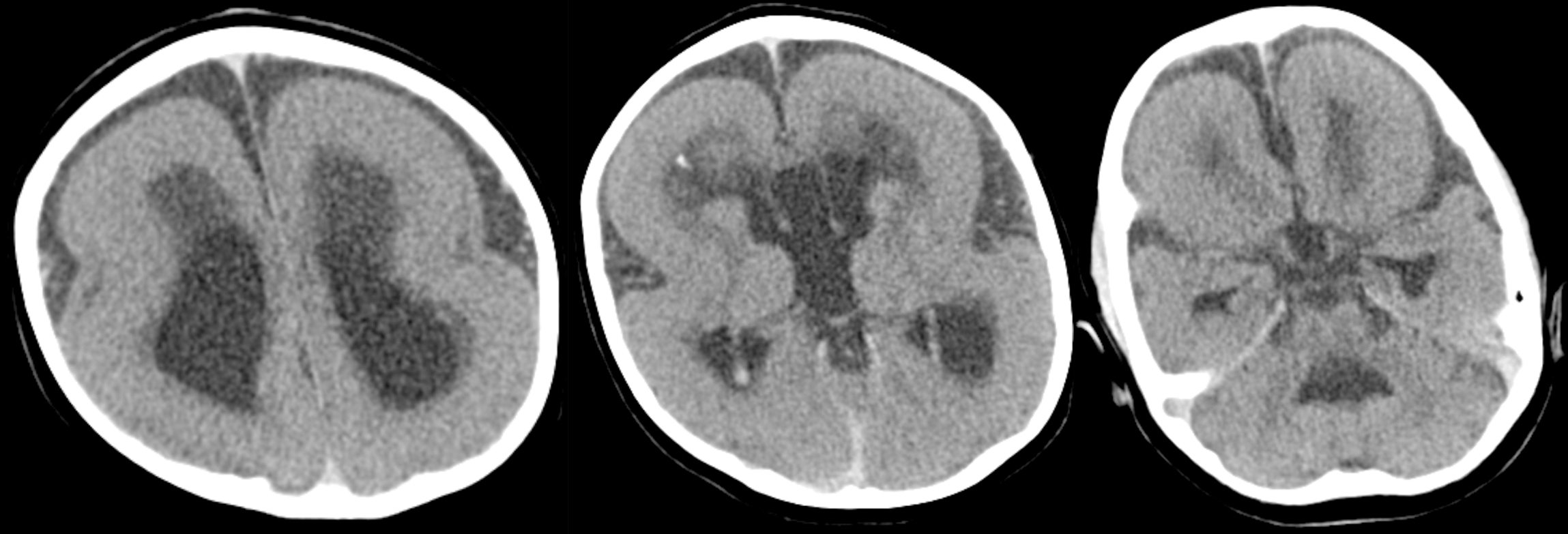
A pediatric patient with a history of prenatal hydrocephalus and epilepsy. 3 axial slices through the brain show a very thickened cortex lacking any of the normal gyrations. The sylvian fissures are broad and shallow. Although difficult to appreciate on these images, the head circumference is small.
PAFAH1B1 (also known as LIS1) on chromosome 17 encodes a protein involved in cellular structure and neuronal migration during early brain development. When this gene is defective, there neurons cannot migrate outward from their origin near the ventricles. The end result is a small brain (microcephaly), a cortex that hugs the ventricular margins (since the neurons never migrated away), with simplified cortical architecture lacking gyrations (agyria). The agyria and thickened cortex (pachygyria) that diffusely affects the brain is what's known as classic lissencephaly, and if microcephaly is also present, then it may also be referred to as microlissencephaly.
An even more severe form involves the deletion of a portion of chromosome 17, such that LIS1 as well as another gene, YWHAE (also involved in neuronal migration), are both lost. This magnifies the findings seen in classic lissencephaly and is known as Miller-Dieker syndrome.
